Lower tray with the tongue in the tongue space
Superseded as the published lower design on 22 September 2026. This page records the 40-mirror tongue-plate study and the evidence behind it. The current lower default is the R24 6 × 6 mm layout lower-tongue-cohort-2lights-24mirrors (mirror positions searched again against six patients’ lower arches on 24 September 2026), which keeps the same tongue plate and tongue proxy; see the smaller handle evaluation.
21 September 2026 · the published lower design since 21 September 2026; the previous lower design is superseded
The previous lower design was searched and evaluated with the tongue space open: nothing sat in it, and its 40 mirrors saw the lingual side of the teeth and gingiva largely from the back of the tray, looking forward across the space where the patient's tongue will be. This study puts an opaque tongue there, counts what that design loses, and redesigns the lower tray around a thin tongue plate that follows the lingual side of the arch and is intended to hold the tongue off the lingual surfaces, with the mirrors re-placed so that the lingual side is captured with the tongue present. All figures are line-of-sight geometric capture at the seated pose with the frozen optical kernel; none is measured hardware performance or reconstruction accuracy. Region names below are the study's geometric proxies, not validated tooth or gingiva labels.
What the tongue does to the previous design
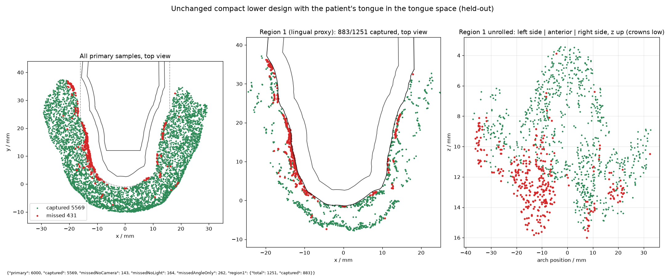
The tongue is a rigid opaque solid. For the unchanged tray it fills the lingual envelope of the arch, the region inside the most lingual scanned points of the teeth and gingiva at any height, shrunk 0.5 mm, through the full height of the tray, out beyond its rear edge as a 44 mm wide slab, minus the tray material. It is not a measured tongue: it does not move over the capture, conform to the crowns or carry saliva. A resting tongue pressed against the lingual surfaces themselves would hide them entirely, so this model is a mild case for the anterior surfaces and a demanding one for the routes that cross the tongue space.
| Held-out capture, 6,000 samples | Previous design, tongue space open | Previous design, tongue present |
|---|---|---|
| All primary surfaces | 5,992 · 99.9% | 5,569 · 92.8% |
| Tray-facing surfaces, region 0 | 2,722 · 100% | 2,683 · 98.6% |
| Inner-facing sides, lingual proxy, region 1 | 1,251 · 100% | 883 · 70.6% |
| Outer-facing sides, region 2 | 2,019 · 99.6% | 2,003 · 98.8% |
| Recessed-surface proxy | 698 of 701 · 99.6% | 676 · 96.4% |
| Two-camera stereo on the lingual proxy | 100% | 69.7% |
The loss sits where a lingual plate has to work: lingual gingiva at z = 9 to 16 mm (326 of 462 samples captured between 9 and 12 mm, 126 of 281 between 12 and 14, 40 of 95 above 14) and the stretch of both arms from y = 2 to 22 mm, where 36 to 62% of each 4 mm band is captured, the left arm faring far worse (15 to 48%) than the right (62 to 84%); the last band, y = 34 to 38 mm, keeps 3 of 31. The anterior lingual surfaces stay covered because the modelled tongue stops at the envelope's front, 0.5 to 0.9 mm behind the most lingual anterior points at gingival height and 3 to 6 mm behind the anterior crowns. Only one held-out sample touches the tongue solid; the loss is blocked routes, not samples buried in the proxy.
The tongue plate
The plate is a 2 mm wall standing 2 mm inside the lingual envelope, measured from the most lingual scanned point at any height, and following the arch all the way round. Along the arms those points are gingiva that leans in over the channel, so the channel between plate and teeth is 2 mm wide at gingiva level and 6 to 9 mm wide at crown level between y = 4 and 28 mm, narrowing to about 5 mm at y = 32 and 2 to 3 mm behind y = 36 mm. The wall rises from the tray's underside to z = 16 mm along the arms, the top of the scanned lingual anatomy (16.3 mm) and 2.3 mm above the tray rim; at the front it stops at z = 14 mm with a 6 mm wide, 3 mm deep midline notch left for the frenum, tapering to full height between y = 8 and 14 mm. Its front face stands at y = 0.6 mm and the tongue space is open right behind it. A floor fill closes the old tongue-space cut between the plate and the tray, and a short bridge on each side from y = 39.3 mm to the tray's rear edge at 41.1 mm closes the channel against the tray's end wall. Everything is one watertight solid with the frozen lower L tray, 27,337 mm³ against 22,193 mm³, with no mesh crossing against the scan and the same 1.36 mm sampled minimum clearance as before. The plate carries no mirror pockets: a 2 mm wall is thinner than a mirror backing, so it is a shield only.
An earlier version of this study, published for a few hours on 21 September 2026, used a 4 mm offset with a 4.5 mm mirror foot and a solid front lobe that left the tongue a chamber only from y = 12 mm backwards, 12 to 26 mm wide. The owner judged that space too narrow and too short, and the design was reworked to the thin plate above; that version captured 5,959 of 6,000 held-out samples and 1,245 of 1,251 on the lingual proxy, so the wider tongue space below costs 25 lingual samples.
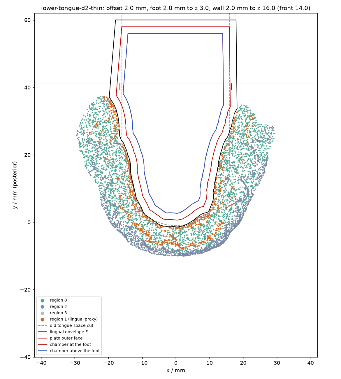
The chamber left for the tongue begins at y = 2.6 mm, just behind the incisors' lingual envelope, and is 8 mm wide at y = 4, 15 mm at y = 8, 16 mm at y = 12, 19 mm at y = 20, 26 mm at y = 30 and 30 mm at the tray's rear edge; the original open tongue space was 32 mm wide. Inside and behind the tray the modelled tongue has 85% of the volume the open tongue space allowed. The plate assumes the tongue can sit slightly further back and higher, as a lingual flange asks of it; the scan contains no floor of mouth, mylohyoid ridge, frenum or sulcus against which to check that. In the redesigned tray the tongue is modelled as the chamber, 0.3 mm clear of the plate, a spill over the plate's top out to 0.5 mm inside the envelope, which brings it within 0.5 mm of the scanned lingual surfaces above the plate, and the same slab beyond the tray's rear edge; nothing is modelled inside the channel.
Search and evaluation
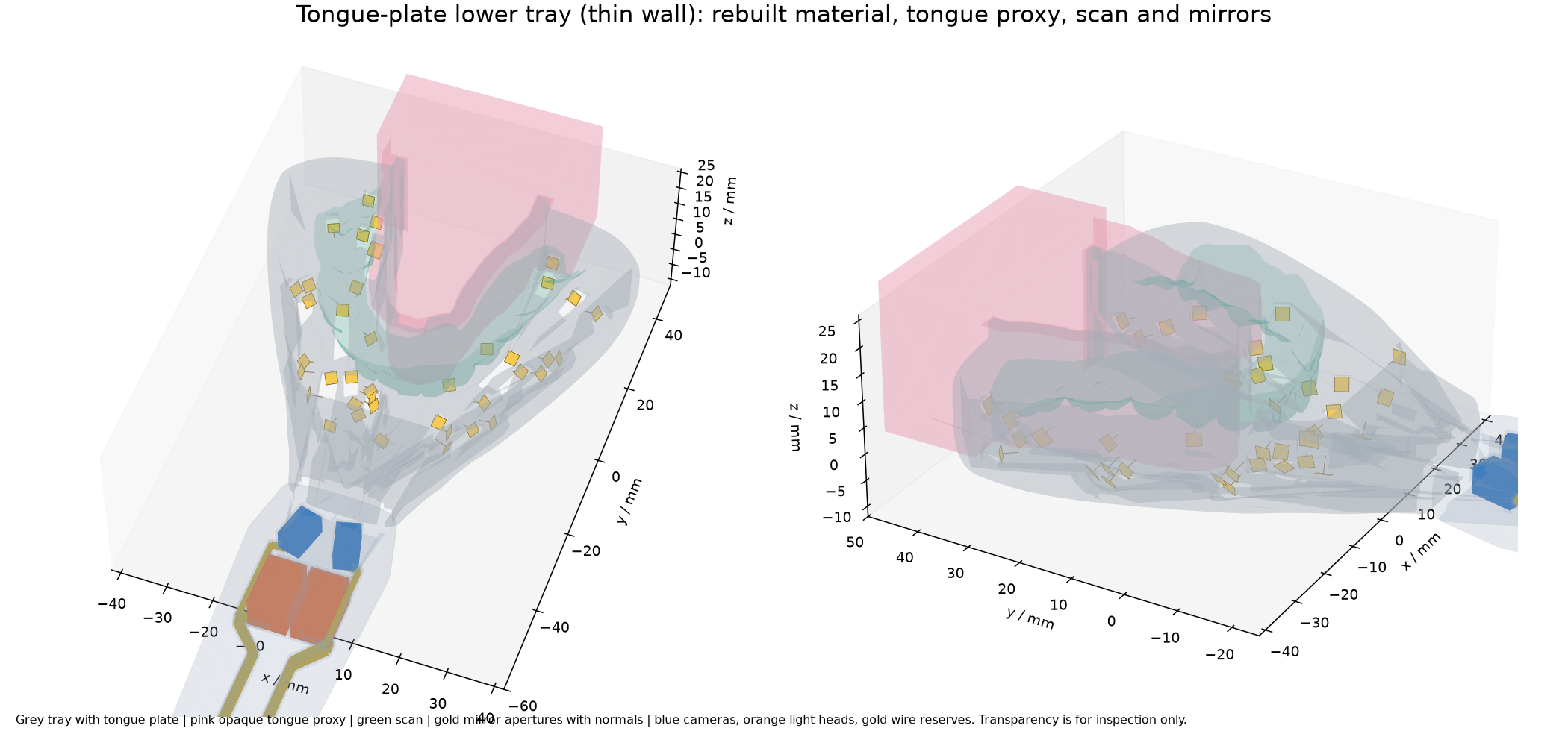
The mirror search is the compact pipeline unchanged in its optics: the same two cameras, two light modules, shared driver and twenty wiring reserves inside the original handle, the same immutable one-reflection kernel, the same mirror forms and the same greedy joint camera-and-light selection. Beyond the geometry, the differences are these. Seats are seeded on the plate tray (754 seeds: walls, rims, floor and the floor fill, none on the plate, with anything inside the chamber or within 0.7 mm of the tongue rejected), and the tongue is an opaque blocker in the search's anatomy, so no feed corridor can be cut through it. The coarse cache held 4,380 candidate mirrors on 500 training points; 1,400 were re-scored on all 3,000 training points with every opaque body present, and the 40-mirror layout was taken from that frontier (screen 99.7%; 24 mirrors already screened at 98.6%). The material was then rebuilt with the 40 finite feed corridors and pockets cut; the project's remaining-seat audit found one floor socket under the anterior teeth left floating by the cuts, so a passive seat bridge of 0.49 mm³ was grown to the nearest material with the project's own script and the sockets recut. The frozen evaluator then traced the result against the actual solids with the tongue as an opaque part of the housing, on the training points and then on the independent held-out set (seed 20260920102), which was not used to shape the plate, the tongue or the layout.
Results
| Held-out capture, 6,000 samples | Previous design, tongue present | Tongue plate, tongue present |
|---|---|---|
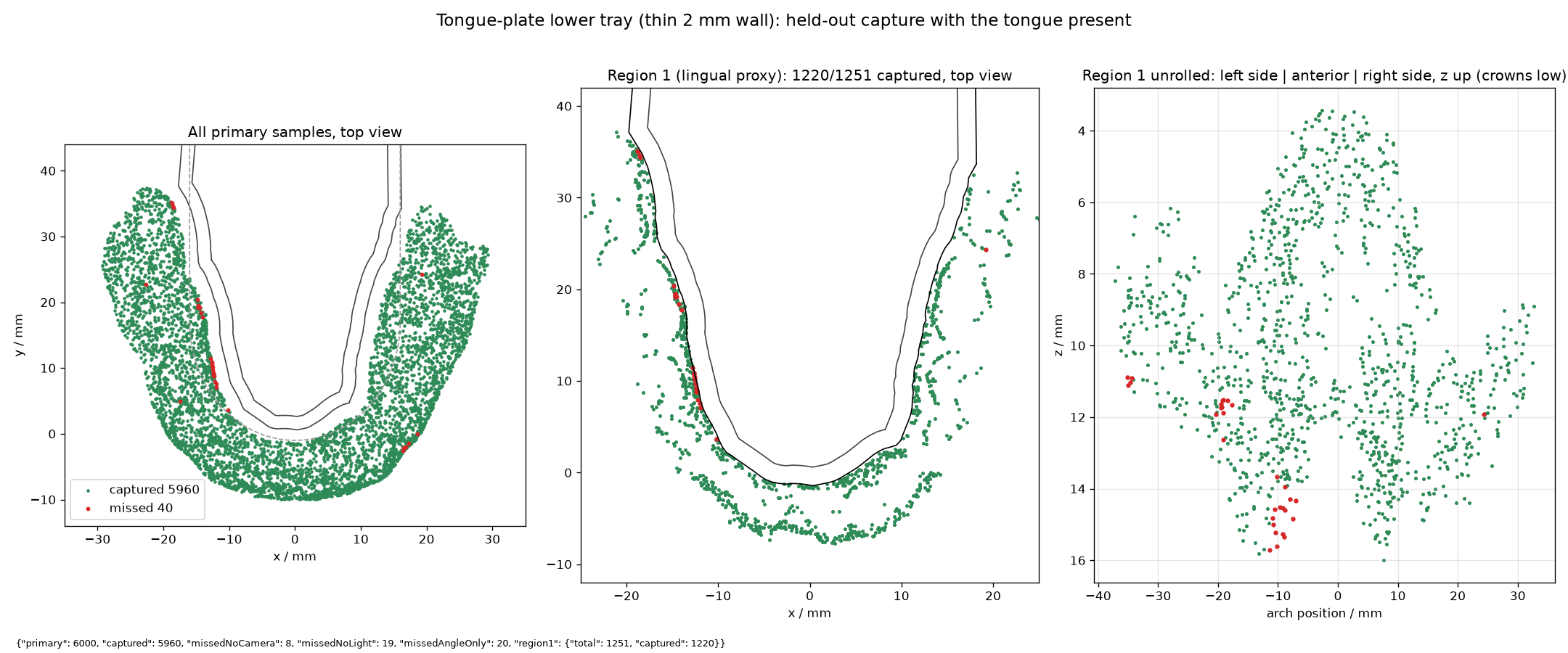
| All primary surfaces | 5,569 · 92.8% | 5,960 · 99.3% (95% sampling interval 99.1–99.5%) |
| Tray-facing surfaces, region 0 | 2,683 · 98.6% | 2,722 · 100% |
| Inner-facing sides, lingual proxy, region 1 | 883 · 70.6% | 1,220 · 97.5% |
| Outer-facing sides, region 2 | 2,003 · 98.8% | 2,018 · 99.6% |
| Recessed-surface proxy | 676 · 96.4% | 698 · 99.6% |
| Two-camera stereo, all · lingual proxy | 92.5% · 69.7% | 99.0% · 97.0% |
| Camera-visible · illuminated | 97.6% · 97.3% | 99.9% · 99.7% |
| Sampling proxy, median · P90 mm per pixel | 2.27 · 4.63 | 2.04 · 3.81 |
Against the previous design with the tongue space open, the plate design captures 5,955 of the same samples, gains 5 and loses 37. Of its 40 misses, 31 are lingual, 30 of them on the left arm at z = 11 to 16 mm, spread from y = 4 to 35 mm, the deep lingual gingiva that the 2 mm gap forces the floor mirrors to see almost edge-on; 12 of them are camera-visible but unlit and 7 are reached by neither leg. The other 9 are outer-facing. On the lingual proxy the anterior bands are complete; the arm bands run from 91 to 99% (6 to 10 mm: 112 of 121; 10 to 14: 98 of 104; 18 to 22: 91 of 100) and the last, y = 34 to 38 mm, keeps 27 of 31. By height the lingual proxy is complete below z = 9 mm, 448 of 462 between 9 and 12, 278 of 281 between 12 and 14 and 81 of 95 above 14. The physical training trace on the rebuilt material reads 2,979 of 3,000 (99.3%) against the search's screening figure of 99.7%.
The lingual surfaces are seen from below. Among captured lingual samples the smallest angle between the surface normal and the arriving camera route has a median of 49° and a 90th percentile of 73°, with 33% beyond 60° and 8% beyond 75°; the previous design with the tongue present manages 48°, 78°, 34% and 13% on the samples it still reaches, and no incidence figure exists for it with the tongue space open. Across all primary samples the median is 31°. Capture at these angles is line of sight, not image quality, and the sampling proxy remains coarse.
Where the mirrors are. All 40 are spherical caps with a 2.2 mm backing: 39 are 2.5 × 2.5 mm (23 of radius 2.4 mm, 16 of 3 mm) and one is 3 × 3 mm with a 5 mm radius, a larger aperture than any in the previous design. Fifteen are fed by camera C1, eight by C2, eight by light L1 and nine by L2. Twenty-seven sit on the tray floor, six on the outer wall, five on the inner face of the front wall and two on the floor fill; none on the plate. Thirty-five of the 40 caps lie below z = 3 mm, the height below which the handle cameras still see under the crowns; the highest is at 10.2 mm on the front wall. The lingual side is captured from the floor, looking up past the crowns to the gingiva that leans in over the channel. Every mirror keeps at least 0.79 mm from the tongue proxy and 1.55 mm from the scan.
Placement and material checks. The rebuilt tray is one connected solid; the exporter discarded 0.007 mm³ of Boolean dust and nothing else. Every feed corridor is a window through the tray's front wall. The chamber wall was probed at 7,247 points a quarter millimetre inside its surface over the full height, the frenum notch excepted: all are material, so at every probe point no feed corridor or pocket reaches the tongue. At one millimetre inside, 6 of 7,246 probes find the wall thinned below 1 mm, all in one spot at the left front at floor level (x ≈ −7.5 mm, y ≈ 5 to 6 mm), where a feed corridor grazes the plate's base; the wall there is between 0.25 and 1 mm thick. The project's remaining-seat audit finds geometric seat material at all 40 sockets with no floating or intruding socket, after the one bridge above. A stricter point-by-point test written for this study gates cap protrusion, tongue and scan clearance and chamber-wall integrity; backing depth and remaining seat material are reported only, because a calibration run on the previous design shows the same pattern under this test. On those measures the new layout is comparable: caps stand up to 1.79 mm proud of the unpocketed tray surface (previous design 1.84 mm), ten mirrors have less than half of their backing inside the unpocketed tray where the floor or front wall is thin (previous design fourteen), and twelve have less than 30% of the material around their pocket remaining (previous design eight). Seat proximity does not establish retention, strength or manufacturability.
What this does not show
The tongue is a static, rigid, opaque proxy; a real tongue moves during the multi-second capture, conforms to the crowns, and could press into the open 2 mm channel or carry saliva onto the mirrors, none of which is modelled. The plate's offset, thickness, heights and notch are design choices bounded by this one scan; two offsets were searched (4 mm with a mirror foot and solid front, then this 2 mm thin plate), nothing in between, and no sensitivity to the wall height or notch was run. The scan ends at the lingual gingiva, so fit against the floor of the mouth, mylohyoid ridge, frenum and sulcus is unverified, and the plate reaches 2.3 mm above the tray rim. Tongue pressure on a wall standing 22 mm tall and 2 mm thick, from the tray's underside to z = 16 mm, and its effect on the stability of a stationary tray, are not assessed. Cross-arch routes for the tray-facing and buccal surfaces also changed and were re-searched, so this is a different layout, not the previous one with additions. Capture is geometric access at one seated pose in the large tray with one anonymised scan; the 20–50 µm accuracy target, structured-light decoding, fluorescence and near-infrared performance remain unaddressed. The upper tray and its published design are untouched and the tongue proxy has no upper counterpart. Since 21 September 2026 this layout is the lower default in the split coverage viewer, where the tongue proxy can be shown or hidden and the published masks include it; the previous lower design is superseded and kept only in the engineering archive.
Files
- Optical layout · housing specification · plate geometry and audits
- Held-out evaluation · held-out summary by band and angle · training evaluation
- Placement audit · remaining-seat audit
- Previous design with the tongue present: evaluation · summary
- Pocketed tray STL, 14 MB · plate material only STL · tongue proxy in the plate STL · tongue proxy in the previous tray STL
The engineering tree holds the full case under tongue-study-assets/lower/lower-tongue-d2-thin/dense/lower-tongue-2lights-40mirrors, the variant geometry under tongue-study-assets/geometry/lower-tongue-d2-thin, the earlier 4 mm variant under lower-tongue-d4, and the scripts that built and checked them.